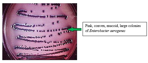
Theory:
Water is unsafe to consume if gas producing lactose fermenting microorganisms present in water. A gas formation may be due to non-coliform bacteria such as Clostridium perfringens, which are gram-positive, and also it may due to gram-negative coliform. The presence of Coliform bacteria is an indication of the unsuitability of water. E.colithat is one of coliform are usually resided in human intestine and known to be fecal in origin. To confirm the presence of gram-negative lactose fermenters in water, the confirmed test is carried out.
Showing posts with label Microbiology Practicals. Show all posts
Showing posts with label Microbiology Practicals. Show all posts
Streak Plate Method
First sufficient amount of agar was poured into a sterile petri dish. Next, this was allowed to solidify. Then a sterilized inoculating needle was dipped in the Durham tube containing test tube. Next, the agar plate was streaked to separate the two types of coliforms. Finally, this was incubated at 370C.
Labels:
Microbiology Practicals
Spread Plate Method
Sufficient amount of agar was poured into a sterile petri dish. Next, this was allowed to solidify. 0.1ml of the fermented water samples were transferred on to the agar surface. A spreader was dipped in 70% alcohol and was flamed. Finally, the sample was spread over the surface of the agar then lids were closed.
Labels:
Microbiology Practicals
Pour Plate Method
First 1ml of the fermented water samples were transferred into sterile Petri dishes, using a sterile pipette. 15ml of Eosin methylene blue agar was poured into the Petri dish and rotate the Petri dish while on the table to mix the water sample with the agar. Next, this was allowed to solidify. Finally, this incubated at 370C.
Labels:
Microbiology Practicals
Most Probable Number (MPN) Test for Water (Viable Cells)
Theory
The routine microbial examination of water to determine its portability is not based on the isolation and identification of pathogenic microorganisms but based on finding a microorganism whose presence indicates that water has been contaminated with an intestinal origin, and therefore the possibility of the presence of pathogenic microorganisms.
The fecal contamination of drinking water is evaluated by using coliform bacteria as an indicator. An indicator organism is an organism that can be used as an indication of pollutions.
Coliforms ferment lactose within 48 hours and produce lactic acid and carbon dioxide.
Lactose --------> Lactic acid + carbon dioxide
In the coliform test, determining the sanitary quality of water involves 4 stages such as;
- Presumptive test
- Confirmed test
- Completed test
- Positive completed test
In this lab presumptive test is carried out. In the presumptive test, the indicator is phenol red. The color changes from red to yellow. In the basic medium, phenol red is red whereas in the acidic medium it is yellow.
Procedure
First two serial dilutions 10-1, 10-2 of water samples provided, were prepared. Next 1ml aliquots were transferred (each from 100, 10-1, 10-2 tubes) into the lactose broth in Durham fermentation tubes. This was done in duplicates. Finally, the tubes were incubated at 350C for 48 hours.
Observation
|
Sample
|
No of positive tubes
|
CFU/ml
|
CFU/100ml
|
|||||
|
100
|
10-1
|
10-2
|
||||||
|
1
|
2
|
1
|
2
|
1
|
2
|
|||
|
Tap water
|
0
|
0
|
0
|
0
|
0
|
0
|
0
|
0
|
|
Tubewell water
|
0
|
1
|
0
|
1
|
0
|
1
|
2
|
2*100 = 200
|
|
Rain water
|
1
|
1
|
1
|
1
|
0
|
1
|
70
|
70*100 =7000
|
|
Bottled water
|
0
|
0
|
0
|
0
|
0
|
0
|
0
|
0
|
|
Well water
|
1
|
1
|
1
|
1
|
0
|
1
|
70
|
70*100 =7000
|
|
River water
|
1
|
1
|
1
|
1
|
1
|
0
|
70
|
70*100 =7000
|
|
Drain water
|
1
|
1
|
1
|
1
|
1
|
1
|
100
|
100*100 =10000
|
|
Stream water
|
1
|
1
|
1
|
1
|
1
|
1
|
100
|
100*100 =10000
|
Discussion
The presence of specific indicator organism signals that a given water sample is contaminated with pathogens. The most widely used indicator for microbial water contamination is the coliform group. Coliforms are used as indicators of water contaminations because many of them inhibit the intestine of humans and other animals in large numbers. Thus their presence in water indicates fecal contamination. Therefore most probable number method can be used to detect and enumerate coliforms in water.
Lactose fermentation after 48 hours at 350C indicates a positive presumptive test for the presence of coliforms in the sample. In this practical, the indicator is phenol red. In a neutral medium, phenol red gives an orange color while it gives a yellow color in acidic medium. Hence the color changes from orange to yellow shows that the medium has changed from neutral to acidic. That indicates the formation of lactic acid due to lactose fermentation. The gas bubble in the fermentation tube also indicates the lactose fermentation. Gas bubble is carbon dioxide. By referring to a statistical table no of coliforms per 100ml can be obtained (MPN Table).
According to the above results, the highest number of colony forming unit (CFU)/100ml was shown by drain water and stream water. Stream water flows through various areas and also polluted water from industries which are coming from drainages used to flow into streams. Hence both drain and stream water are highly polluted. Thus the coliform contaminations may be higher, due to the exposure to various situations. Next highest is well water, river water, and rainwater. Well water does not flow. River water flows but does not expose to various areas as the stream water. This is why CFU of river water does not exceed the CFU of stream water. Although rainwater is exposed it does not contaminate with pollutants. But CFU value of rainwater has a high value. It may be due to instrumental or experimental errors. This can occur due to unclean containers or when counting a deformed gas bubble may be counted. Because when the gas bubble is very large the bottom of the gas bubble is difficult to identify. Both tap and bottled water have zero values, which indicates these two samples are not contaminated with coliforms. Since tap water is purified before distributing to houses it is already checked for coliforms. Only if it is free from coliforms the distribution is done. Bottled water is also purified and sterilized.
For the success of the results, it is important to use sterile containers to collect water samples. And a wide-mouthed, sterile, glass-stoppered bottles can be used. When collecting tap water, let the water to flow initially and then take the sample, whereas, in a stream, water is collected below the water surface. If transportation is a necessary water sample is packed in ice. Otherwise, due to environmental conditions, the activity of the organism may change. To inhibit further activities of coliforms, after 48 hours the samples should place in a refrigerator.
According to the national water and drainage board, total coliforms equal to 10 while fecal coliforms equal to 0, are acceptable values in drinking water /100ml. These values can vary depending on the country.
Labels:
Microbiology Practicals
IMViC Test
Theory:
This is a group of tests used particularly in the identification of the family Enterobacteriaceae. This group consists of Escherichia coli, Salmonella sp, Shigella sp, Enterobacter sp, Proteus sp, Eruinia sp.
IMViC test contains several tests such as;
- I = Indole test
- M = Methyl red test
- V = Voges Proskauer test
- C = Citrate utilization test
Indole test carried out in sim agar media;
Methyl red test carried out in MR-VP Broth;
Reaction with E.coli;
E.coli can change the pH of the medium into an amount of 4.
Reaction with E.aerogenes;
E.aerogenes can change the pH of the medium into an amount of 6.5.
Voges Proskauer test carried out in MR-VP Broth;
This changes the pH around 6.0.
Citrate test carried out in Simmons citrate medium;
Color change in Indole test is from pale yellow to red and methyl red test pale yellow to red. In Voges Proskauer test is from yellow to red and in citrate test, it is from green to blue.
Procedure:
Indole Test
First, the colonies of E.coli and E.aerogenes from the completed test were used to inoculate with tryptone broth in separate test tubes. Next, these were incubated at 370C for 2-5 days. Finally, the indole was tested by Kovac’s method.
Kovac’s reagent was added carefully to the test medium dropwise to form a layer at the top of the medium. Finally, the tube was gently agitated with a rotary motion.
Kovac’s reagent: 5g of para dimethylamino benzaldehyde dissolved in a mixture of 75ml of amyl alcohol and 25ml of concentrated HCl.
Methyl red Test
First, the colonies of E.coli and E.aerogenes from the completed test were used to inoculate with MR-VP (dextrose phosphate medium) in separate test tubes. And this was incubated at 370C for 48 hours. Finally, a few drops of an alcoholic solution of methyl red were added to the tube.
Voges Proskauer Test
First, the colonies of E.coli and E.aerogenes from the completed test were used to inoculate with MR-VP (dextrose phosphate medium) in separate test tubes. And this was incubated at 370C for 2-5 days. In order to test Acetylmethylcarbinol, 0.5ml of naphthol solution (5% alcoholic) and 0.5ml of 40% KOH containing 0.3% creatine were added to about 3ml of the test medium. Next, the tubes were stoppered and shaken vigorously and were placed in a slanted position for 5-15 minutes.
Citrate Utilization Test
First, the colonies of E.coli and E.aerogenes from the completed test were used to inoculate with Koser’s citrate medium. And Bromothymol blue was added as an indicator.
Observations:
|
Test
|
Observation
|
Conclusion
|
|
|
Indole Test
|
A red ring was formed on top of the peptone solution which was incubated with E.coli and the pale yellowish color of the solution remained the same.
No such formation of a red ring has occurred in the broth which was incubated with E.aerogenes.
|
E.coli present.
|
|
|
Methyl red Test
|
MR-VP medium with E.coli was formed a red ring on top of the solution and color of the solution remained the same.
MR-VP medium with E.aerogenes has not formed a red ring.
|
E.coli present.
|
|
|
Voges Proskauer Test
|
MR-VP medium with E.aerogenes turned the yellow solution into dark red whereas no specific change in MR-VP medium with E.coli.
|
E.aerogenes present.
|
|
|
Citrate Utilization Test
|
Koser’s citrate medium with E.aerogenes turned the green solution into blue whereas no change in color in Koser’s citrate medium with E.coli
|
E.aerogenes present.
|
|
Discussion:
In order to provide definite biochemical proof that the isolated organism is indeed E.coli a series of test, usually referred to as the IMViC tests are used. These can be used both to characterize E.coli and to differentiate it from Enterobacter aerogenes. Colonies from the completed test used to inoculate several different media. And this test can be used to identify other species in family Enterobacteriaceae.
A peptone medium rich in the amino acid tryptophan is inoculated and allowed to grow for 24 hours. E.coli makes the enzyme tryptophanase which forms indole, pyruvic acid, and ammonia from tryptophan. Since E.aerogenes cannot catabolize tryptophan, testing for the presence of indole differentiate these two organisms. Formation of red ring indicates the presence of E.coli.
P-dimethyl aminobenz-aldehyde + Indole ------> Cherry-red compound
Methyl red is an acid-base indicator that turns red in a slightly acidic medium. Thus if methyl red is added to a culture medium containing glucose in which an organism has grown for 18 to 24 hours, a red color will be observed which would indicate that organic acids had been formed as a result of fermentation of the glucose. E.coli forms large amounts of acids, which make methyl red positive. E.aerogenes on the other hand, carries out a 2,3-butylene glycol type of fermentation and thus produces only small amounts of organic acids which makes methyl red negative.
The Voges Proskauer test detects the presence of acetoin (Acetylmethylcarbinol), the immediate precursor of 2, 3-butylene glycol fermentation which is positive for E.aerogenes and negative for E.coli and the positive result is indicated by a pink ring.
Acetyl methyl carbonyl + Napthol ------> Diacetyl +Guanidine (group of peptone)
This reaction is carried out in the 40% KOH oxidation. The pink color is obtained both by Diacetyl and guanidine.
Citrate test merely determines whether the organism in question can grow using citrate as its sole source of carbon. Although both E.coli and E.aerogenes possess the necessary enzymes to metabolize citrate, only E.aerogenes can use it as a carbon source. E.coli cannot use citrate because citrate cannot enter the cells of E.coli. This test, therefore, determines the difference in the ability of these two organisms to transport citrate across their cell membranes.
Citrase
Citric acid -------> Oxalic acid + acetic acid + pyruvic acid + CO2 excess
2CO2 + 2 Na+ + 2 H2O ----> Na2CO3 ----> Colour change from green to blue
Hope this article helpful for your lab practical and please share this among your friends.
Labels:
Microbiology Practicals
Subscribe to:
Posts (Atom)
Popular Posts
-
Gram stain is a method of differential staining. Gram stain was discovered and introduced by Christian Gram in 1884. This divides bacteria ...
-
The main causation for the spoilage of food is by microbes. There are three types of microorganisms that cause food spoilage. Those are bact...
-
Nuts are regularly prone to chemical changes. Fats and oils are the major constituents of nuts. Rancidity causes due to high content of oil ...
-
There are various culture media. Mixed culture, pure culture and in vitro ( cultured in containers), in vivo (grown in live animal and plan...
-
Theory: Water is unsafe to consume if gas producing lactose fermenting microorganisms present in water. A gas formation may be due to non-...
-
Physical damages on food items is caused through wounds, Stress conditions such as water stress, nutrimental stresses etc., extreme conditio...
-
Theory The routine microbial examination of water to determine its portability is not based on the isolation and identification of path...
-
Observation of living cells follows below procedures. Negative Staining Microorganism will not stain in this method, but the medium...
-
Theory: This is a group of tests used particularly in the identification of the family Enterobacteriaceae. This group consists of Escher...
-
Microorganisms are invisible to the naked eye. Therefore for the purpose of studying, they are cultured. Staining will kill microorganism...
Microbiology Basics
Microbiology Tests
Food Microbiology
- Effect of Interactions on Food Microbes
- Food Spoilage
- Microbial Activity on Food Spoilage
- Chemical Changes on Food Spoilage
- Effect of Physical Damage on Food Spoilage
- Freezer Burn in Food Spoilage
- Staling Effect for Food Spoilage
- Contaminating Micro Flora Composition Factors
- Factors Determining the Composition of the Contaminating Micro Flora
- Effect of Nutrient Compositionon Food Microbes
- Spoilage of Canned Foods in Microbiology Aspect


